
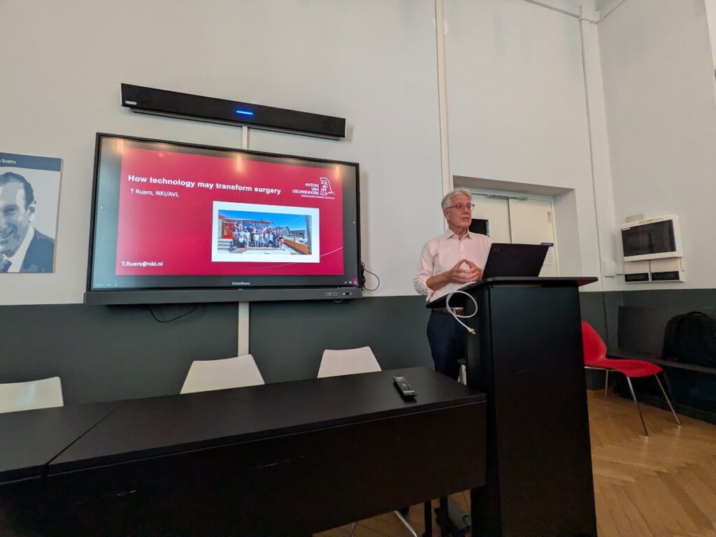
On 21 July 2026, SpectraBREAST launched its webinar series with Theo Ruers, surgical oncologist at the Netherlands Cancer Institute, and his talk How Technology May Transform Surgery. The hybrid event was hosted at the Department of Physics of Politecnico di Milano and attended both in person and online.
It was a great pleasure to welcome Theo Ruers to Milan and learn from his unique experience at the intersection of surgery, biomedical imaging and technology.
“I am first and foremost a surgeon.”
This clinical perspective shaped the central message of the talk: technology should not be developed simply because it is technically possible, but because it addresses a real surgical need.
Surgery still needs better ways to see
Although surgeons have access to detailed imaging before an operation, once in the operating room they still rely mainly on sight and touch.
“When you’re operating, you have to rely on your feelings and on your vision. And that’s really not that much.”
Ruers compared an easily identifiable tumour to a marble inside a sponge. In reality, however, a tumour is often more like “a sponge in a sponge”, with no clear boundary separating malignant and healthy tissue.
One of the most urgent needs is therefore to determine during surgery whether tumour tissue remains at the resection margin. Conventional pathology may provide this information only several days later, when the patient may already require a second procedure.
“If you tell the surgeon during surgery, they can easily remove an additional part.”
Ruers presented optical approaches including diffuse reflectance spectroscopy and hyperspectral imaging, showing their potential to distinguish tumour from healthy tissue and provide rapid guidance during open, laparoscopic and robotic surgery.
The challenge of previously treated tumours
Ruers placed particular emphasis on patients who receive chemotherapy before surgery. After treatment, the tumour may shrink or break up into small residual areas, making it even harder for the surgeon to recognise its true extent.
“It becomes even more difficult to find, see and feel the tumour. Sometimes it shrinks and sometimes it disintegrates. Then you have all these small spots.”
Technologies for surgical guidance must therefore be validated not only on clearly visible tumours, but also in patients whose tissue and tumour morphology have been substantially altered by treatment.
The opposite situation creates an equally important challenge: some patients achieve a complete response, meaning that no residual tumour remains after therapy. However, current imaging methods cannot always establish this with sufficient certainty.
“We have no technology at this stage to determine this complete response. MRI doesn’t work, CT doesn’t work, PET doesn’t work.”
As a result, some patients may still undergo surgery even though the tumour has disappeared. Developing optical methods capable of detecting minimal residual disease—or reliably confirming its absence—could therefore improve surgical precision and potentially avoid unnecessary procedures.
From technical performance to clinical adoption
A strong message from the webinar was that performance alone is not enough. New technologies must be fast, compact, affordable, simple to operate and seamlessly integrated into the surgical workflow.
“If it makes their workflow very complicated, they will never use it.”
“Your technology should be fast—in five or six minutes, I would say.”
These practical requirements determine whether an innovation can move from the laboratory into routine patient care.
The webinar concluded with a lively discussion on clinical validation, usability, measurement depth and pathology correlation. It was an excellent start to the SpectraBREAST webinar series, which will continue with further events dedicated to hyperspectral imaging, cancer research and clinical translation.